Xoilac Tv chắc hẳn là trang web quen thuộc với những người hâm mộ thể thao. Bởi sân chơi hàng đầu này luôn đồng hành cùng anh em qua các mùa giải trong và ngoài nước.
Xoilac Tv là website phát trực tiếp bóng đá miễn phí đỉnh cao hàng đầu tại thị trường Việt Nam. Nơi đây cung cấp vô vàn tính năng hữu ích khác nhau giúp người dùng nhanh chóng tìm kiếm, cũng như xem thông tin mà mình cần. Cùng khám phá nhé!
Tổng quan về Xoilac Tv
Ngay khi nhận thấy nhu cầu xem trực tiếp bóng đá tại Việt Nam ngày càng tăng lên. Tuy nhiên, vào thời điểm đó, các trang web phát bóng đá vẫn chưa thể đáp ứng được đầy đủ mọi nhu cầu của khách hàng. Dàn chuyên gia hàng đầu đã ngay lập tức hợp tác với các kỹ thuật viên IT tài năng để tạo nên kênh Xoilac Tv.
Website được thành lập với mục tiêu là nơi xem bóng đá số 1 thị trường Việt Nam nên sự đầu tư khá chuyên nghiệp và bài bản. Từ việc mua bản quyền phát sóng những giải đấu lớn cho đến ứng dụng công nghệ hiện đại. Qua đó, gián tiếp mang đến vô vàn trận đấu hấp dẫn chất lượng cao để bất cứ ai cũng có thể thoải mái tận hưởng miễn phí.

Vì thế, chỉ trong một khoảng thời gian ngắn, Xoilac Tv đã phát triển nhanh chóng đến bất ngờ để hiện tại trở thành địa chỉ được nhiều anh em lựa chọn. Nếu muốn xem bất kỳ trận bóng đá trực tiếp diễn ra trên điện thoại hoặc máy tính hằng ngày thì giờ đã là việc quá đơn giản,
Ngoài phát sóng bóng đá trực tiếp, hệ thống còn sở hữu thêm nhiều tính năng khác. Khi sử dụng, người chơi có thể tìm và xem những thông tin liên quan đến thể thao mình mong muốn dễ dàng. Tất nhiên, mọi nguồn dữ liệu đều đầy đủ và chính xác.
Những tính năng chính trên trang chủ Xoilac Tv
Xoilac Tv mang đầy đủ những ưu điểm cần thiết của trang live bóng đá hàng đầu hiện nay, cụ thể:
Trực tiếp bóng đá
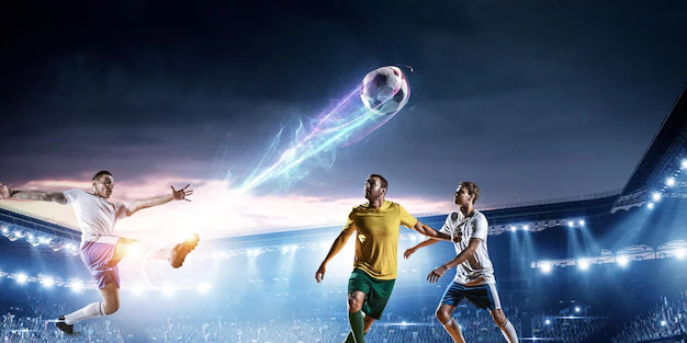
Là nền tảng truyền hình trực tiếp các trận bóng đá hàng đầu thế giới. Khi đến với Xoilac Tv, người xem có thể tận hưởng những phút giây thăng hoa ngay trên màn hình full HD. Từ diễn biến chính đến những pha bóng kịch tính và kết quả cuối cùng, mọi thứ đều được truyền tải chân thực và vô cùng sống động.

Những thông tin quan trọng sẽ không bao giờ bị bỏ lỡ, khán giả dễ dàng theo dõi diễn biến trận đấu và cảm nhận được hơi thở từ sân cỏ. Xoilac Tv đồng hành cùng người hâm mộ bóng đá, mang đến nhiều trải nghiệm tuyệt vời và thú vị giống như đang ngồi ngay trên khán đài của sân vận động.
Lịch thi đấu mới nhất
Ngoài việc phát trực tiếp các trận đấu thì Xoilac Tv còn cung cấp lịch thi cụ thể các giải bóng đá trong nước lẫn quốc tế, giúp người xem thu thập thông tin dễ dàng. Thông qua trang web, fan hâm mộ có thể nhanh chóng nắm chính xác thời gian, địa điểm cũng như những thông tin quan trọng liên quan đến trận đấu. Từ đó, anh em tiện sắp xếp, lên kế hoạch theo dõi hợp lý nhất.
Nhờ sự tiện lợi này, Xoilac Tv không chỉ là dịch vụ truyền thông bình thường mà còn là nguồn tài nguyên quan trọng để mọi người sử dụng thời gian được hiệu quả, thú vị hơn, đồng thời tận hưởng niềm đam mê của mình.
Bình luận, phân tích chi tiết trận đấu
Xoilac Tv đã tạo ra nền tảng thú vị dành cho người hâm mộ túc cầu, cung cấp cho họ các phân tích chi tiết và ý nghĩa về trận đấu. Bằng việc cung cấp những video phân tích chất lượng cao, Xoilac cho phép người xem của mình nắm bắt được những khoảnh khắc quan trọng của mỗi trận đấu, từ phối hợp chiến thuật đến những pha bóng quyết định. Điều này góp phần tạo ra trải nghiệm độc đáo mang tính giáo dục.
Sự góp mặt của các bình luận viên, chuyên gia bóng đá là điểm nhấn tạo nên sự khác biệt cho Xoilac. Với kiến thức sâu rộng của mình, họ đưa ra những bình luận sâu sắc về mọi khía cạnh của trận cầu. Từ bước phân tích chiến thuật, nhận xét về màn trình diễn của cầu thủ hay bình luận về sự kiện quan trọng, giúp người xem tiếp cận thực tế từ một góc nhìn hoàn toàn mới,
Tương tác với người xem
Xoilac Tv không đơn thuần là nền tảng xem bóng đá trực tiếp mà còn mở ra cơ hội cho anh em tham gia vào cộng đồng cùng chung đam mê. Tại đây, thành viên vừa được xem trực tiếp, lại vừa được tham gia thảo luận, bày tỏ quan điểm, chia sẻ cảm xúc với bạn bè bốn phương trên toàn thế giới.
Nhờ đó, môi trường tương tác trở nên sống động hơn bao giờ hết, giúp kết nối người hâm mộ túc cầu lại với nhau. Việc tạo ra không gian giao lưu, chia sẻ ngoài làm phong phú trải nghiệm xem bóng đá, mà còn thể hiện sức mạnh to lớn của niềm đam mê thể thao trong việc gắn kết con người lại với nhau.
Đánh giá ưu điểm của Xoilac Tv trên thị trường hiện nay
Có thể nói, trên thị trường hiện nay xuất hiện rất nhiều trang cá cược thể thao phục vụ nhu cầu giải trí của thị trường. Tuy nhiên, ưu điểm của Xoilac Tv là một trong những đơn vị tiên phong trong lĩnh vực bóng đá. Ngoài ra, hệ thống còn tạo ấn tượng nhờ kinh nghiệm lâu năm, chắc chắn sẽ không làm người xem thất vọng.
Phát sóng đa dạng các mùa giải
Là website thể thao bóng đá hàng đầu giúp fan hâm mộ tự do lựa chọn đồng hành với số lượng giải phong phú, đa dạng. Tất cả các trận đấu bóng đá trực tuyến đều được tổng hợp tại đây, kể cả những sân chơi có bản quyền.
Ngoài ra, các trận đấu khi phát sóng đều được xếp theo thứ tự, không chỉ gói gọn trong khu vực Đông Nam Á hay châu Á mà Xoilac Tv còn live cả những trận diễn ra trên toàn thế giới. Vì thế, nó tạo ra sự phấn khích và hài lòng cho đa số người xem.
Giao diện cực thân thiện
Với mục đích giúp người chơi thoải mái tìm kiếm thông tin cầu thủ, trận đấu hay bất cứ dữ liệu thể thao nào. Vì vậy, website đã tạo ra một giao diện trang chủ mà tất cả thành viên đều có thể sử dụng được ngay từ lần đầu.
Hình ảnh sắc nét, chỉnh chu, kiến thức cập nhật rõ ràng, các chuyên mục được đặt logic,… Mỗi danh mục sẽ chứa một vấn đề khác nhau, đảm bảo không lẫn lộn với các nguồn khác.
Thiết kế chất lượng cao
Nếu đến đây để giải trí, chắc chắn người chơi sẽ có những trải nghiệm vô cùng tuyệt vời. Hệ thống trang chủ Xoilac Tv luôn được duy trì và đầu tư cẩn thận về chất lượng cũng như hình ảnh, âm thanh sắc nét đến từng chi tiết.

Link trận đấu sẽ được hệ thống cập nhật cho anh em sớm nhất là trước 1h trước khi bóng lăn. Ngoài ra, buổi phát sóng trực tiếp được trình chiếu với độ phân giải cao cùng dàn âm thanh hấp dẫn, sôi động, mang đến cho người xem cảm giác như hòa mình vào không khí ở khán đài. Hơn nữa, tốc độ truyền tải mượt mà không có sự chậm trễ.
Xem bóng đá miễn phí
Ưu điểm cực lớn của Xoilac Tv là khán giả có thể xem bóng đá hoàn toàn miễn phí. Điều này đã mang lại sự tiện lợi cũng như tiết kiệm chi phí cho những ai không muốn tiêu tiền quá nhiều. Hệ thống cung cấp một vài kênh free, giúp người xem thoải mái truy cập mà không lo ảnh hưởng vốn.
Đường truyền mượt mà
Được đánh giá cao về tốc độ khi truyền tải. Hệ thống mạng của Xoilac Tv phải tối ưu hóa nhằm đảm bảo cho người xem mãn nhãn mà không lo gián đoạn hay giật lag. Điều này vô cùng quan trọng khiến thành viên có thể thưởng thức trận đấu mà không bị phân tâm khi xem.
Tỷ lệ kèo hấp dẫn
Tại đây, đơn vị không chỉ tập trung vào phát sóng các trận đấu bóng đá với nhiều loại cược đa dạng. Ngoài ra, website Xoilac còn đưa ra tỷ lệ khác nhau nhằm đáp nhu cầu như: kèo châu Á, kèo châu Âu, kèo tài xỉu, kèo phạt góc,… Vậy nên, càng làm tăng sự phấn khích của thành viên.
Hướng dẫn cụ thể xem bóng đá Xoilac Tv trực tiếp
Như đã đề cập ở trên, website Xoilac được thiết kế rất khoa học và chia thành nhiều phần khác nhau. Ngoài ra, sân chơi còn tương thích với một số hệ điều hành cho điện thoại, laptop, máy tính,… Vì vậy, nếu muốn tìm và xem bất cứ màn chạm trán nào tại đây cũng vô cùng đơn giản. Chỉ cần thực hiện:
- Bước 1: Đầu tiên, anh em truy cập vào các trình duyệt quen thuộc như Google Chrome, Cốc Cốc, Firefox, Opera,…
- Bước 2: Vào phần tìm kiếm trên website, nhập từ khóa liên quan như “bóng đá trực tiếp xoilac”, “Xoilac Tv”, “xem bóng đá tại Xoilac”,…
- Bước 3: Truy cập địa chỉ chính thống bên dưới phần tìm kết quả.
- Bước 4: Tiếp đến, chọn Trang chủ và kéo xuống. Tại đây, người chơi có thể xem tất cả các link trực tiếp bóng đá hay nhất trong ngày.
- Bước 5: Cuối cùng, cược thủ chỉ cần chọn trò chơi mình muốn thưởng thức.
Website Xoilac Tv được thiết kế rất khoa học
Vài lưu ý cần nhớ khi xem bóng đá Xoilac Tv
Để có trải nghiệm xem bóng đá trên Xoilac tốt nhất hiện nay, người chơi nên lưu ý những thông tin quan trọng sau:
- Chuẩn bị thiết bị có kết nối internet thật ổn định để giúp quá trình phát trực tiếp diễn ra suôn sẻ mà không sợ tình trạng giật, lag.
- Nếu gặp tình trạng bị đơ, treo máy,…khi đang xem thì có thể do trục trặc từ thiết bị nào đó hoặc cấu hình của máy quá thấp không đủ đáp ứng được nhu cầu. Anh em nên tạm dừng khoảng 1-2 phút, nhấn F5 để làm mới và tải lại trang để khắc phục tình trạng.

- Hệ thống Xoilac Tv luôn cung cấp link dự phòng cũng như liên kết của các server khác để giảm tải cho link chính. Thế nên, người xem hoàn toàn có thể tiến hành chuyển đổi sang đường link bên cạnh của mỗi trận khi cần.
- Để mang về trải nghiệm xem bóng đá tốt nhất, dàn nhân viên bình luận của Xoilac Tv luôn là những người nhiệt tình truyền lửa cho khán giả. Anh em cũng nên mời bạn bè, người thân theo dõi bóng đá trên kênh mọi lúc mọi nơi để chia sẻ niềm vui bất tận.
Kết luận
Sứ mệnh của Xoilac Tv là tạo ra một không gian tương tác kết hợp thông tin đa dạng về mảng bóng đá. Từ đó, tạo sự gắn kết, phấn khích trong cộng đồng fan hâm mộ. Đây thực sự là một địa chỉ hấp dẫn và chất lượng, đảm bảo sẽ mang đến cho người xem phút giây thư giãn cực kỳ tuyệt vời.